Automated Segmentation of Post-Surgical Resection Cavities on MRI - a MELD study
1 in 5 epilepsy patients experience seizures caused by structural abnormality in the brain. For these patients, resection surgery to remove the epileptogenic zone is one of the best chances of cure, and complete resection of the epileptogenic zone is important for achieving seizure freedom. Quantitative assessment of resection completeness requires:
- accurate delineation of the postoperative resection cavity, and
- comparison with the preoperative lesion
However, existing delineation approaches are often time-consuming, labour-intensive, and difficult to generalise across datasets and imaging protocols.
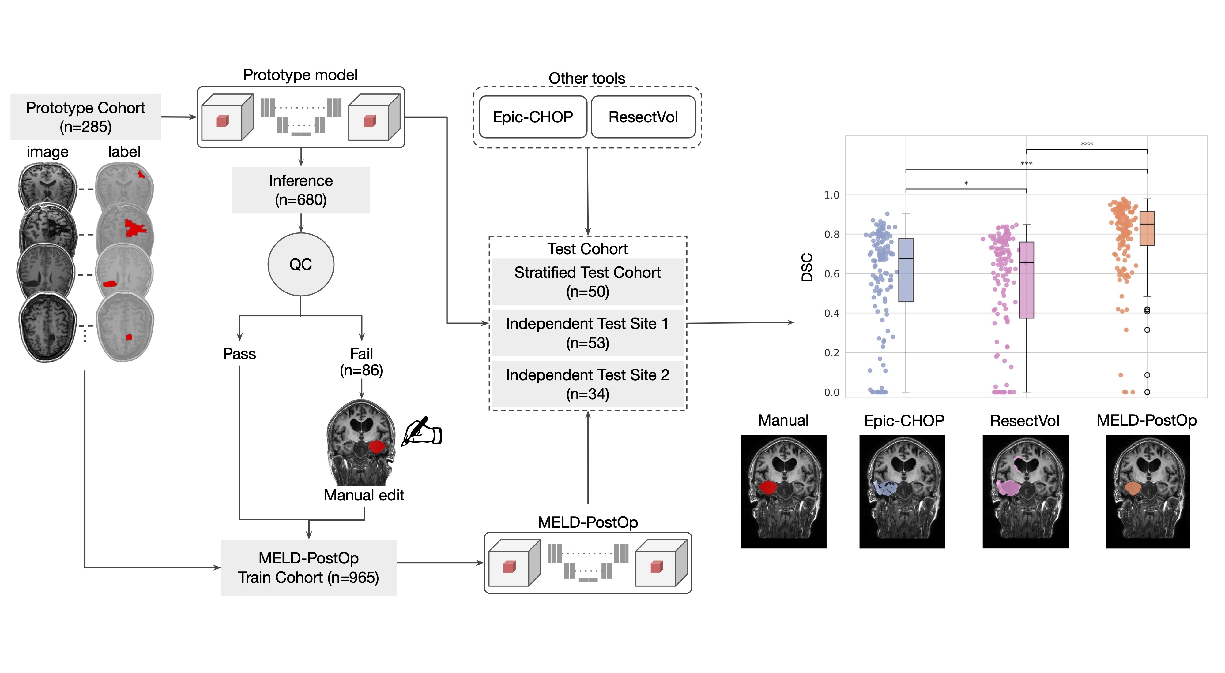
To address this challenge, we developed MELD-PosTOp, a deep learning tool that automatically segments resection caivites directly from postoperative 3D T1-weighted MRI scans, without requiring additional preprocessing or additional modalities.
The model was trained using 965 postoperative scans with annotations, sourced from the MELD Focal Epilepsies dataset and the open-source EPISURG dataset
Results
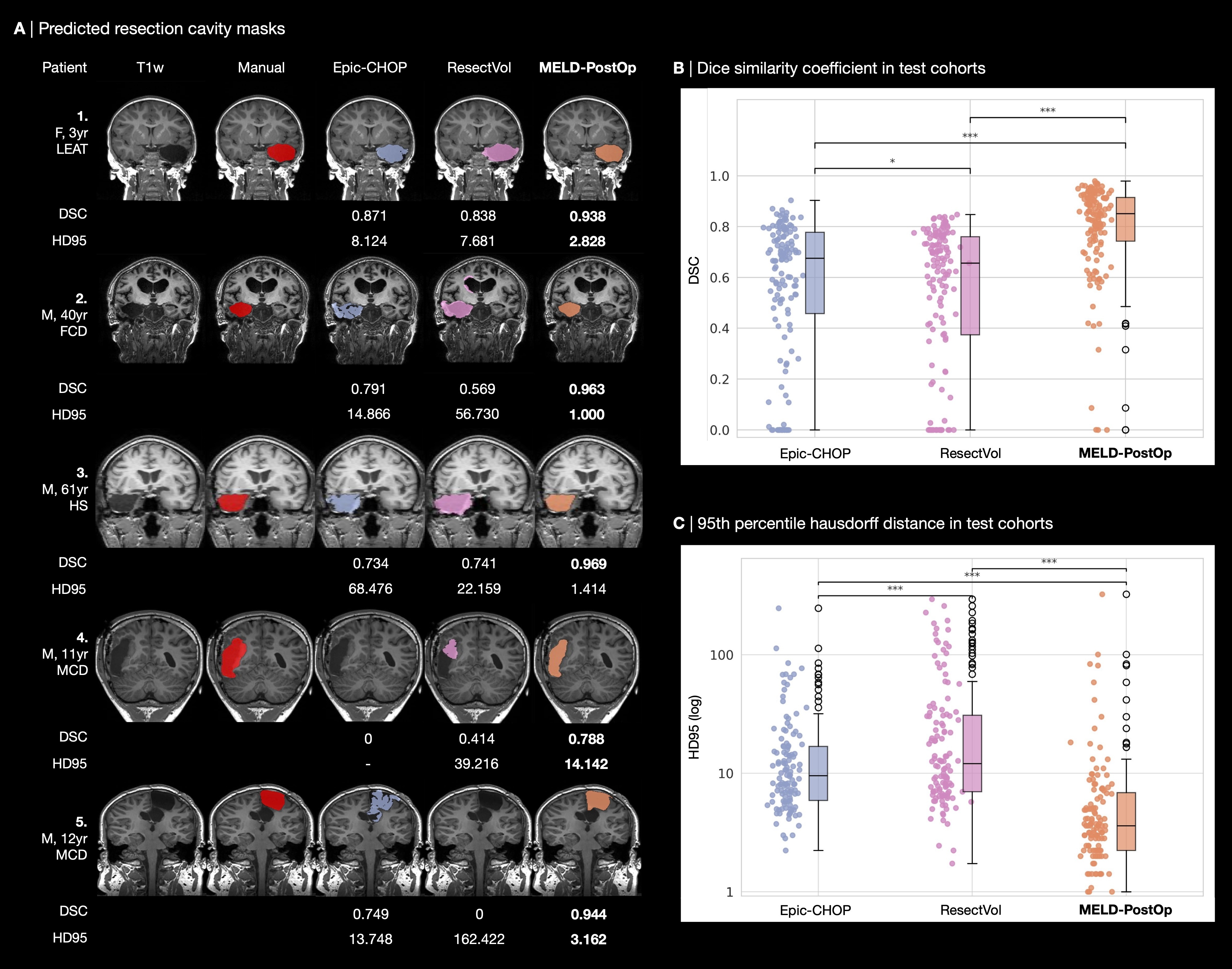
MELD-PostOp demonstrates fast, reproducible, and generalisable segmentation performance across large and heterogeneous imaging cohorts. MELD-PostOp detected 135/137 resection cavities (98.5%), and 128 segmentations achieved meaningful overlap with ground truth segmentation (Dice > 0.5). No statistically significant performance differences were shown across: sex, age (adult vs paediatric), pathology (HS vs non-HS), surgical lobe (temporal vs extra-temporal), surgical side, MRI field strength, and image isotropy. Runtime was 17 seconds per image, compared to >10 minutes for other tools.
Installation & Usage
Instructions for installation and usage can be found here: MELD-PostOp Github Page
Pre-trained model weights can be downloaded here: MELD-PostOp Figshare Page
Preprint
For more details, preprint is available here: MELD-PostOp MedRxiv